Midface and Zygoma Fracture

Midface and Zygoma Fracture

Module Summary

The surgical treatment goal in repairing midface fractures is to restore masticatory function and orbital function and to restore facial form. Restoration of facial buttresses and dental occlusion restores function and facial width and projection. Restoration of orbital volume and release of incarcerated orbital tissues restores orbital form and function. Midface fractures require a comprehensive timing and treatment plan for each fracture. Lateral canthotomy with inferior cantholysis is a quick way to decompress an orbit involved in midface trauma. ZMC and orbit are considered together during repair and should be best managed when swelling has subsided. Soft tissue replacement following surgical access is essential to prevent esthetic complications.  
 

Module Learning Objectives 

After completing this module the physician will be able to:

  1. Discuss the initial assessment and management of life-threatening and orbit-threatening issues often seen with severe midface face trauma.  
  2. Identify the boundaries of the bones of the midface to include the maxilla, zygoma, nasal bones, orbit bones, frontal bone and pterygoid bones. 
  3. Discuss the structural bony anatomy of the midface and describe its system of vertical and horizontal buttress.   
  4. Describe the anatomy of the zygoma and the articulations associated with complex fractures of the zygoma.  
  5. Describe the common injury patterns of midface fractures and include the LeFort classification, zygomaticomaxillary (ZMC) fractures and orbital fractures.  
  6. Diagnose midface and zygoma fractures by patient symptoms, physical exam presentation, and radiographic findings.
  7. Accurately identify fractures using CT and 3DCT radiographic findings to prescribe the appropriate treatment required for midface and zygoma fractures.  
  8. Explain the soft tissue approaches for surgical repair of the midface.    
  9. Describe the repair sequence of complex panfacial trauma.   
  10. Explain the dental and occlusal considerations in midface fractures.   
  11. Discuss the surgical technique necessary to reconstruct the three-dimensional skeletal structures to restore the face to its original width, height, and sagittal projection and occlusion.  
  12. Describe complications associated with midface and zygoma fractures and discuss treatment for bleeding, malunion, nerve injury, eye and lacrimal injury, and malocclusion. 

Anatomy

Learning Objectives 
  1. Name and identify the bones that make up the midface, include the frontal, nasal, maxillary, palatal, orbital, and zygomatic regions.
  2. Identify the bony articulation of the maxilla and zygoma with the nose, orbit and cranium.  
  3. Identify the four attachments of the zygoma to surrounding bones.
  4. Discuss the neural structures that course through the orbit and identify the path of the infraorbital nerve from posterior to anterior in the floor of the orbit.  
  5. Identify the vertical and horizontal buttresses of the midface and orbits.  
References 
  1. Agur AMR, Dalley AF. Grant's Atlas of Anatomy. 14th ed. Baltimore: Lippincott Williams & Wilkins; 2016.  
  2. Kellman RM. Maxillofacial Trauma. In: Flint et al. Cummings Otolaryngology: Head and Neck Surgery. 6th ed. Philadelphia, PA: Mosby Elsevier; 2014. p. 326-331.  
  3. Song WC, Choi HG, Kim SH, et al. Topographic anatomy of the zygomatic arch and temporal fossa: A cadaveric study. J Plast Reconstr Aesthet Surg. 2009;62:1375-1378.
  4. Patterson R. The Le Fort fractures: René Le Fort and his work in anatomical pathology. Can J Surg. 1991;34:183–184.

Pathogenesis

Learning Objectives 
  1. Discuss the biomechanics associated with midface and zygoma fractures.  
  2. Discuss the protective effect of the neuro-cranium by the facial “crumple zone”. 
  3. Describe the Le Fort midface fracture classification.
  4. Describe what causes increased orbital volume in zygomatic fractures.
References 
  1. Patterson R. The Le Fort fractures: René Le Fort and his work in anatomical pathology. Can J Surg. 1991;34:183–184.
  2. Hopper RA, Salemy S, Sze RW: Diagnosis of midface fractures with CT: what the surgeon needs to know. Radiographics. 2006;26:783–793.
  3. Kellman RM. Maxillofacial Trauma.  In: Flint et al. Cummings Otolaryngology: Head and Neck Surgery. 6th ed. Philadelphia, PA: Mosby Elsevier; 2014. p. 327-330.  
  4. Kühnel TS, Reichert TE.  Trauma of the midface.  GMS Curr Top Otorhinolaryngol Head Neck Surg. 2015; 14: Published online 2015 Dec 22. 

Incidence

Learning Objectives 
  1. Discuss the three most common midface fractures.  
  2. Describe the most common causes of midface fractures.  
  3. Describe the associated ocular injuries with midface fractures.
References 
  1. Septa D, Newaskar VP, Agrawal D, Tibra S. Etiology, Incidence and Patterns of Mid-Face Fractures and Associated Ocular Injuries.  J Maxillofac Oral Surg. 2014;13:115-119.
  2. Lee EI, Mohan K, Koshy JC, Hollier LH:  Optimizing the Surgical Management of Zygomaticomaxillary Complex Fractures. Semin Plast Surg. 2010;24: 389–397.

Patient Evaluation

Learning Objectives 
  1. Discuss a thorough physical examination to include airway, visual, vascular, and neurological systems in patients with midface fractures. 
  2. Describe the bony and soft tissue findings of the cheek and orbit in the zygomaticomaxillary complex (ZMC) fractures.
  3. Describe the position of the maxilla in examination of a patient with a mobile Le Fort fracture and include the occlusal relationships that can exist in this setting.    
  4. Midface trauma may be associated with life-threatening and sight-threatening complications, discuss priorities in the systematic and repeated assessment required in overall care of these patients. 
References 
  1. Lee EI, Mohan K, Koshy JC, Hollier LH:  Optimizing the Surgical Management of Zygomaticomaxillary Complex Fractures. Semin Plast Surg. 2010;24:389–397. 
  2. Kellman RM. Maxillofacial Trauma.  In: Flint et al. Cummings Otolaryngology: Head and Neck Surgery. 6th ed. Philadelphia, PA: Mosby Elsevier; 2014:331-332 
  3. Nastri AL, Gurney B: Current concepts in midface fracture management.  Current Opinion in Otolaryngology & Head and Neck Surgery.  2016; 24:368–375.
  4. Moe KS (Jun 17, 2018). Maxillary and Le Fort Fractures Treatment & Management.  Medscape. Retrieved September 30, 2018, from https://emedicine.medscape.com/article/1283568-overview.
  5. Resident Manual of Trauma to the Face, Head, and Neck. (2012) First Edition.  AAO-HNS Foundation.  Retrieved August 24, 2018, from https://www.entnet.org/sites/default/files/ResidentTraumaFINALhighres.pdf.

Imaging

Learning Objectives 
  1. Cite which imaging study is best to evaluate most midface and zygoma fractures?
  2. Discuss the best imaging technique to view the orbital apex and optic canal.  
  3. Discuss the imaging technique that would best aid the surgeon in treatment planning zygomatic fractures that involve complex three-dimensional position changes as well as involvement of the lateral and inferior orbital walls.  
  4. Identify what imaging modality is best suited for interoperative bony repositioning.  
References 
  1. Hopper RA, Salemy S, Sze RW: Diagnosis of midface fractures with CT: what the surgeon needs to know. Radiographics. 2006;26:783–793.
  2. Winegar BA, Murillo H, Tantiwongkosi B:  Spectrum of critical imaging findings in complex facial skeletal trauma. Radiographics : a review publication of the Radiological Society of North America, Inc. 2013;33:3–19. 
  3. Kellman RM. Maxillofacial Trauma.  In: Flint et al. Cummings Otolaryngology: Head and Neck Surgery. 6th ed. Philadelphia, PA: Mosby Elsevier; 2014. p. 332-333. 

Treatment

Learning Objectives 
  1. Discuss the importance of delaying facial fracture repair until the appropriate consultations have been made and the patient has been stabilized.      
  2. Identify the need for antibiotic treatment or prophylaxis prior to surgical access.  
  3. Discuss the importance of a good physical exam and axial and coronal CT to establish a well-designed treatment plan for appropriate exposure and repair sequencing.  
  4. Describe how the design and repair of unstable segments becomes more complex when treating panfacial fractures.  
     
References 
  1. Winegar BA, Murillo H, Tantiwongkosi B:  Spectrum of critical imaging findings in complex facial skeletal trauma. Radiographics : a review publication of the Radiological Society of North America, Inc. 2013;33:3–19. 
  2. Kellman RM. Maxillofacial Trauma.  In: Flint et al. Cummings Otolaryngology: Head and Neck Surgery. 6th ed. Philadelphia, PA: Mosby Elsevier; 2014. p. 341-349.  
  3. Curtis W, Horswell B B. Panfacial fractures: an approach to management. Oral Maxillofac Surg Clin North Am. 2013;25:649–660.  
  4. Martou G, Antonyshyn O M. Advances in surgical approaches to the upper facial skeleton. Curr Opin Otolaryngol Head Neck Surg. 2011;19:242–247.  

Pharmacology

Learning Objectives 
  1. Describe the role of antibiotics in the treatment of facial fractures.
  2. Discuss surgical wound healing and the use of steroids in midface fracture repair.  
References 
  1. Zosa BM, Elliott CW, Kurlander DE, et al.  Facing the facts on prophylactic antibiotics for facial fractures: 1 day or less.  J Trauma Acute Care Surg. 2018;85:444-450.
  2. Soong PL, Schaller B, Zix J, Iizuka T, Mottini M, Lieger O. The role of postoperative prophylactic antibiotics in the treatment of facial fractures: a randomised, double-blind, placebo-controlled pilot clinical study. Part 3: Le Fort and zygomatic fractures in 94 patients. Br J Oral Maxillofac Surg. 2014;52:329-333.
  3. Snäll J, Kormi E, Koivusalo AM, Lindqvist C, Suominen AL, Törnwall J, et al. Effects of perioperatively administered dexamethasone on surgical wound healing in patients undergoing surgery for zygomatic fracture: a prospective study. Oral Surg Oral Med Oral Pathol Oral Radiol. 2014;117:685-689. 

  4.  

Surgical Therapies

Learning Objectives 
  1. Describe the various surgical soft tissue approaches to the upper facial skeleton. 
  2. Describe the difference in treatment of a minimally displaced ZMC fracture compared to one with significant rotation.  
  3. Discuss the importance of the zygomaticosphenoid (ZS) suture in complex ZMC fractures.  
  4. Discuss the reconstruction of the horizontal and vertical buttresses of the Le Fort I through III fracture patterns.  
  5. Discuss the precise relocation of bony subunits and resuspension of soft tissues in achieving acceptable functional and aesthetic outcomes.
  6. Discuss the various types and application of reconstructive methods that are used in the midface.   
  7. Discuss correct plating choices for the maxillary buttresses and infraorbital rims.   
References 
  1. Cornelius CP, Gellrich N, Hillerup S, et al.  Midface Trauma. AO Surgery Reference.  https://www2.aofoundation.org/wps/portal/surgery?showPage=diagnosis&bone....  
  2. Kellman RM. Maxillofacial Trauma.  In: Flint et al. Cummings Otolaryngology: Head and Neck Surgery. 6th ed. Philadelphia, PA: Mosby Elsevier; 2014:341-349.  
  3. Moe KS (Jun 17, 2018). Maxillary and Le Fort Fractures Treatment & Management.  Medscape. Retrieved September 30, 2018, from https://emedicine.medscape.com/article/1283568-overview
  4. Manson PN. Craniofacial Fractures. In: Prien J, editor.  Manual of Internal Fixation in the Cranio-Facial Skeleton.  Springer-Verlag Berlin Heidelberg New York; 1998.  p. 95-138
  5. Lee EI, Mohan K, Koshy JC, Hollier LH:  Optimizing the Surgical Management of Zygomaticomaxillary Complex Fractures. Semin Plast Surg. 2010;24:389–397. 
  6. Curtis W, Horswell B B. Panfacial fractures: an approach to management. Oral Maxillofac Surg Clin North Am. 2013;25:649–660.
  7. Winegar BA, Murillo H, Tantiwongkosi B:  Spectrum of critical imaging findings in complex facial skeletal trauma. Radiographics : a review publication of the Radiological Society of North America, Inc. 2013;33:3–19.

Case Studies

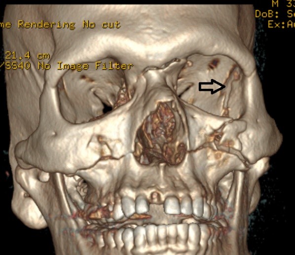
Case 1: ZMC fracture with displaced ZS suture. A 33 y.o. male presented to the ED with a history of club-to-face assault. He was intoxicated with LOC. Upon presentation the ED noted increased left IOP and a lateral canthotomy was performed.  He was then evaluated and cleared by ophthalmology. On examination he denied blurry or double vision but reports malocclusion.  He denied salty taste in mouth but does report bilaterally intermittent clear rhinorrhea with walking. Physical examination was significant for a mobile and retruded maxilla and a depressed left zygoma. The left eye was swollen and decompressed by canthotomy. CT angiogram of the neck showed no evidence of blunt carotid or vertebral artery injury. CT and 3DCT review identified small left temporal lobe hemorrhagic contusions, extensive complex facial/orbital fractures. Left LeFort I, II, III and left zygomatic fracture. Right LeFort I and II fracture.  Hard palate fracture with extension to the alveolar ridge and traumatically absent right central maxillary incisor tooth. Left medial nasal lacrimal duct fracture. Nasal bone and nasal septal fractures. Cribriform plate fractures. Thickening of the left inferior and possible left lateral rectus muscles may suggest entrapment.  
Discussion: Reduction of the maxilla was aided by placing IMF screws anteriorly on the intact mandible and posteriorly on the maxilla allowing maxillary advancement with MMF wire tightening. Transoral vestibular and left transconjunctival approaches were made to access the midfacial skeleton and orbit.  The left ZS suture was accessed through the lateral canthotomy and separate blepharoplasty incision to help orient the zygoma during plating.  A displaced zygomaticosphenoid (ZS) suture (left photo) is seen in the left orbit.  A post-operative scan (right photo) shows the midface repair.  When available, intraoperative CT scanning can be a very helpful in positioning the zygoma. A separate horizontal incision was made over the nasion for the reduction and fixation of the nasofrontal fracture.  Following facial access and MMF the midface was plated and orbital floor repaired.   


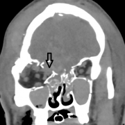
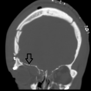
Case 2: Right forehead trauma producing Lefort I-III and brain herniation into right eye. A 25 year old male was transferred from a community hospital where he underwent urgent decompressive craniotomy for right sided EDH. He arrived at our trauma center with a GCS of 3 after being therapeutically paralyzed and intubated. He was stabilized and placed in the SICU and initially sedated on Propofol and placed on Keppra prophylaxis and plan for craniotomy with neurosurgery.  Bilateral chemosis was found by ophthalmology with concern for left globe rupture. There was no evidence of ocular muscle entrapment. The only acute management issues were intubation for acute respiratory failure.  CT head showed severely comminuted fractures involving the frontal bones, orbits and mid face with moderate to large bilateral frontal contusions. Comminuted bilateral orbital roof fractures with herniation of a small amount of brain into the right orbit (left photo).  Fractures involved the left foramen ovale, left foramen spinosum, left carotid canal and right optic canal. Fractures involve the cribriform plates, ethmoid sinuses and sphenoid sinus with marked displacement of fracture fragments. Left temporal bone fracture was seen with ossicular disruption. Diffuse brain swelling with compression of third and lateral ventricles and basilar cisterns was noted and there was no evidence of cervical spine fracture or dislocation.
Discussion: The first considerations are acute management of the EDH and concern for ocular injury. CSF leak would be highly suspected. The midface fractures were produced from the force delivered at the right frontal cranium which is unusual.  His life-threatening issues required close cooperation with neurosurgery.  We delayed the cranial part of the procedure and took the patient in a staged fashion for tracheostomy, MMF and right zygomaticofrontal suture repair. The initial stage would allow for brain rest in anticipation for treating the cranium, cribriform and orbit. The second surgery was done 10 days later. The right photo shows a porous polyethylene/titanium implant placed to separate orbit and brain.  The cribriform area was severely traumatized and was covered with temporalis fascia to prevent CSF leak.  As expected the patient has not regained olfactory function and has not experienced CSF rhinorrhea. Vision and EOM’s were intact.  

References 
  1. Lee EI, Mohan K, Koshy JC, Hollier LH:  Optimizing the Surgical Management of Zygomaticomaxillary Complex Fractures. Semin Plast Surg. 2010;24:389–397.
  2. Cornelius CP, Gellrich N, Hillerup S, et al.  Midface Trauma. AO Surgery Reference.  https://www2.aofoundation.org/wps/portal/surgery?showPage=diagnosis&bone....  
     

Complications

Learning Objectives 
  1. Discuss the problems associated with inadequate reduction of maxillary fractures.  
  2. Describe the complications of imprecise orbital reconstruction.  
  3. Discuss eyelid and globe malposition causes.
  4. Discuss the causes of post-operative diplopia.
  5. Describe dental complications to include tooth injury and malocclusion.
  6. Discuss causes and sites of CSF leak when treating midface trauma.    
  7. Discuss wound infection prevention in midface trauma surgery.  
  8. Discuss prevention and treatment of the lacrimal collecting system when damaged.
  9. Discuss the prevention of unfavorable facial scar formation in midfacial soft tissue following trauma or surgery. 
  10. Discuss proper resuspension of the soft tissues before wound closure.  
  11.  Discuss methods of preventing nerve, brain and ocular injuries during surgery.  
References 
  1. Kellman RM. Maxillofacial Trauma.  In: Flint et al. Cummings Otolaryngology: Head and Neck Surgery. 6th ed. Philadelphia, PA: Mosby Elsevier; 2014:349.  
  2. Moe KS (Jun 17, 2018). Maxillary and Le Fort Fractures Treatment & Management.  Medscape. Retrieved September 30, 2018, from https://emedicine.medscape.com/article/1283568-overview
  3. Hasheminia D, Kalantar Motamedi MR, Hashemzehi H, Nazeri R, Movahedian B. A 7-year study of 1,278 patients with maxillofacial trauma and cerebrospinal fluid leak. J Maxillofac Oral Surg. 2015;14:258-62. 
  4. Ord RA. Post-operative retrobulbar haemorrhage and blindness complicating trauma surgery. Br J Oral Surg. 1981;19:202-207. 
  5. Colletti G, Valassina D, Rabbiosi D, Pedrazzoli M, et al. Traumatic and iatrogenic retrobulbar hemorrhage: an 8-patient series. J Oral Maxillofac Surg. 2012;70:e464-468.
  6. Kelts G, Maturo S, Couch ME, Schmalbach CE. Blunt cerebrovascular injury following craniomaxillofacial fractures: a systematic review. Laryngoscope. 2017;127:79-86.
     

Review

Review Questions 
  1. What are the physical signs and symptoms of zygoma (ZMC) fracture?  
  2. What is the classification for midface fractures?
  3. What bony structures require repair in Le Fort I fractures?
  4. Restoring what middle third facial structure is important for reestablishing the correct facial architecture?
  5. What are the sites of CSF leak in severe facial trauma?   
  6. Where are the three horizontal buttresses located?  
  7. What bone fracture is necessary for a Le Fort fracture? 
  8. What posterior orbit bone stops the Le Fort III fracture from continuing into the optic canal?  
  9. What surgical errors are responsible for malunion and malocclusion?  
  10. What is the benefit of the Carol-Gerard screw?   
  11. How is enophthalmos treated?  
  12. What does a Frost stich prevent?